Cone Beam Imaging: The Future of Dental Imaging Technology
Roam Dental in Shelby Township uses in-office Cone Beam CT (CBCT) 3D imaging to plan implants, extractions, and complex treatments with submillimeter accuracy — no outside imaging center, no referral, no extra appointment. Dr. Virgil Barbu and our team rely on CBCT to visualize teeth, jawbone, nerves, and sinuses from every angle before recommending treatment, so patients from Shelby Township, Utica, Sterling Heights, and across Macomb County get faster diagnoses and safer outcomes. CBCT is essential for dental implant placement, wisdom tooth evaluation, TMJ diagnosis, and root canal planning. Schedule a consultation or call (586) 500-7647 to see how 3D imaging improves your care.
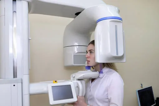
Cone beam computed tomography (CT) is a specialized x-ray machine used when regular dental or facial x-rays are insufficient. While not used routinely due to increased radiation exposure compared to regular dental x-rays, cone beam CT generates highly detailed 3-D images of dental structures, soft tissues, nerve paths, and bone in the craniofacial region in a single scan. This allows for precise treatment planning, similar to conventional CT imaging.
Unlike conventional CT, dental cone beam CT uses a smaller, less expensive machine that can be placed in an outpatient office. While cone beam CT provides detailed images of the bone, it is not as effective as conventional CT in evaluating soft tissue structures such as muscles, lymph nodes, glands, and nerves. However, it does offer lower radiation exposure, making it a safer option for patients.
Dental cone beam CT is commonly employed to aid in the treatment planning of various dental issues. It is especially useful in more complex cases that involve:
Preparation for a cone beam CT examination is simple and straightforward.
Before the procedure, you will need to remove any metal objects that may interfere with the imaging, including jewelry, eyeglasses, hairpins, and hearing aids. While removable dental work may also need to be taken out, it is recommended to bring them to your examination as your dentist or oral surgeon may need to examine them.
For female patients, it's crucial to inform your dentist or oral surgeon if there is any chance that you might be pregnant. Otherwise, there is no special preparation required for the examination.
Advantages:
Risks:
CBCT 3D imaging is reserved for cases where 2D x-rays cannot provide enough information for safe treatment. For implants near the inferior alveolar nerve, impacted wisdom teeth, TMJ evaluation, and complex pathology, the 3D detail prevents surgical errors that would be far more costly to correct later.
At Roam Dental, low-dose settings and smaller fields of view are used whenever clinically appropriate — especially for children — to minimize radiation exposure while maintaining diagnostic quality.
Reviewed by Dr. Virgil Barbu, DDS · Roam Dental, Shelby Township, MI
Advanced CBCT technology provides diagnostic capabilities that traditional x-rays simply cannot match:
See teeth, bone, nerves, and sinuses from every angle — far more diagnostic information than conventional 2D x-rays can provide.
Our CBCT unit is designed for dental use and delivers a fraction of the radiation of a medical CT scan, making it safe for regular diagnostic use.
your dentist uses 3D bone maps to place implants in the optimal position, avoiding nerves and sinuses for safer surgery and better long-term results.
CBCT reveals cysts, infections, root fractures, and bone loss that are invisible on standard x-rays — enabling earlier intervention and better outcomes.
No referral to an outside imaging center. Your scan is performed, processed, and reviewed with your dentist at the same appointment — saving you time.
CBCT is recommended when a 2D x-ray cannot provide enough information for safe treatment planning. Dr. Barbu orders a cone beam scan when the diagnosis, surgical plan, or patient safety depends on seeing bone volume, nerve position, or root anatomy in three dimensions.
You may benefit from CBCT if you are considering:
CBCT may not be needed for: routine cleanings, single-tooth cavities, or straightforward fillings — standard digital x-rays are sufficient in those cases.
3D Imaging is a good fit if you are:
This may not be the right option if:
Here is exactly what happens at your appointment at Roam Dental. Knowing each step makes the experience far less intimidating — and lets you relax knowing what comes next.
Here is how 3D Imaging compares to the main alternatives. Dr. Barbu reviews all options with every patient at Roam Dental so you make the best-informed decision for your situation.
| Feature | 2D Digital X-Ray | CBCT 3D Imaging |
|---|---|---|
| Dimensions captured | 2D (flat) | 3D (volumetric) |
| Radiation dose | Very low | Low |
| Bone volume measurement | Estimate only | Precise (submillimeter) |
| Nerve position mapping | Not visible | Clearly visible |
| Scan time | Seconds | 10–40 seconds |
| Best use | Routine cavities, gum levels | Implants, impactions, TMJ, pathology |
These are the questions patients at Roam Dental ask most often before committing to treatment. Dr. Barbu addresses each one honestly at your consultation.
"CBCT sounds expensive and unnecessary for routine dentistry"
CBCT is only ordered when the diagnostic benefit justifies the cost and exposure — never for a routine cleaning. For implants, extractions near nerves, and unexplained jaw pain, the 3D detail prevents surgical mistakes that would cost far more to fix.
"I'm worried about radiation exposure"
A dental CBCT delivers a fraction of the radiation of a medical CT scan — often comparable to a few days of natural background radiation. Dr. Barbu uses the lowest effective dose and smaller field of view whenever possible.
"Can't a regular x-ray tell you the same thing?"
For simple cases, yes. But 2D x-rays only show two dimensions, so bone depth, nerve position, and root curvature are invisible. CBCT reveals all three dimensions — critical when a millimeter matters.
Every procedure has tradeoffs, and transparent conversation about them is part of informed consent. Dr. Barbu reviews these at your consultation and answers every question before treatment begins.
CBCT 3D imaging provides a much more detailed view of a patient's teeth, jaws, and facial structures than traditional dental X-rays. It produces high-resolution, three-dimensional images that can be viewed from any angle.
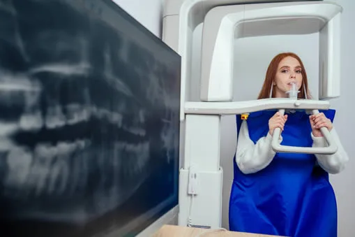
No, CBCT 3D imaging is a painless procedure. The patient simply needs to sit or lie still while the machine rotates around their head.
A CBCT 3D imaging procedure typically takes between 10 and 40 seconds, depending on the area being imaged.
Yes, children can have CBCT 3D imaging. However, the dose of radiation used is lower for children than for adults.
Yes. Roam Dental in Shelby Township offers in-office CBCT 3D imaging to patients across Shelby Township, Utica, Sterling Heights, Clinton Township, and Macomb County. Schedule an appointment or call (586) 500-7647.
Many dental insurance plans cover CBCT scans when they are medically necessary for treatment planning — such as implant placement, impacted teeth, or surgical evaluation. Coverage varies by plan. Our team will verify your benefits before the scan and discuss any out-of-pocket cost during your consultation.
A dental CBCT scan delivers significantly less radiation than a medical CT scan — often comparable to a few days of natural background radiation from the environment. Dr. Barbu orders CBCT only when the diagnostic benefit outweighs the radiation exposure, and low-dose settings are used for children and smaller fields of view.
Yes. If you are referred to a specialist or want a second opinion, we can provide your CBCT images on a CD, USB, or through a secure digital transfer. Just let our front desk know and we will prepare the files for you.
Medical Disclaimer
This content is for informational purposes only and does not constitute dental or medical advice, diagnosis, or treatment. CBCT 3D imaging involves low-dose radiation exposure and should only be performed when the diagnostic benefit outweighs the risk, as determined by your dentist. Consult Dr. Virgil Barbu or a qualified dental professional regarding any questions about your oral health. Individual results may vary.
Contact our office to learn how cone beam CT 3D imaging can improve your diagnosis and treatment planning.